The Collaborative Imaging Platform for Modern Eye Care

See the whole patient story

Unified automatically brings images from every instrument, across all your locations, into one place. View, compare and share them from any computer or your phone.
Search for a patient...
Cmd K
Tag Stats
12.2k User
12.3k Location
847 Organization
3.2k Date
Patient Inbox
Anderson, James
Thompson, Lisa
Garcia, Maria
Brown, David
Wilson, Amy
OD FundusOS Fundus OD Fundus
OCT Macula OCT Macula
1,100+
Locations connected
3,000+
ECP members
4,500+
Devices integrated
15M+
Patient visits unified

Compare visits, side by side

Put today's scan next to last year's, even from a different device.

OD
OD OCT
OCT
OS
OS Fundus
Fundus
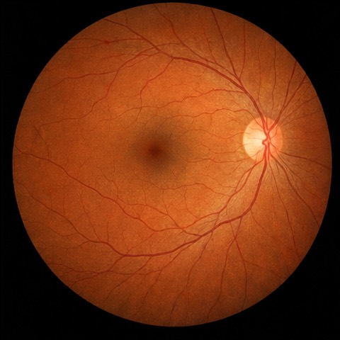
Fundus photograph showing diabetic retinopathy findings
Compare Images
Fundus photograph - baseline
OD Jul 16, 2025 NW400
Fundus photograph - comparison
OD Jan 10, 2025 maestro
Differences
Changes detected: 2 regions
Temporal
Inferior arcade

AI that works with you

AI that flags what to look at and drafts your notes, plus custom models trained on your own images. You make the call.

Retina.help
AI
Color fundus photograph
OD Fundus - Photography
DR Classification
No DR
92.00%
Mild NPDR
8.00%
Moderate NPDR
0.00%
Severe NPDR
0.00%
Virtual Scribe
AI
Color fundus photograph, diabetic retinopathy
Source Image OD Fundus - Photography
Mild non-proliferative diabetic retinopathywith scattered microaneurysms in the posteriorpole. No macular edema detected.
Follow-up in 3 months
Continue current management
Repeat OCT at next visit
AI Confidence: High
Reviewed & Approved
Custom Models
Beta
Color fundus photograph
OD Fundus — Photography
Predictions
drusen
0%
no drusen
100%
Model Performance
Accuracy 94.2%
Training data 1,247 images
Last updated Jan 15, 2026
Available Models

Collaborate in real time

Share a case in seconds with a colleague, a referral, your patient or your research cohort, with HIPAA compliance built in. No more CDs, faxes or email.

Secure Message
HIPAA
Re: Anderson, James — OCT Review
SC
Dr. Chen 2:15 PM
I've reviewed the latest OCT. The macular thickness looks stable compared to October.
2:18 PM Dr. Park
Good to hear. Any concerns about the peripheral findings?
RP
SC
Dr. Chen 2:22 PM
Minor drusen noted temporally OD, but no progression. I'd recommend standard follow-up in 6 months.
OCT_macula_OD.jpg 2.4 MB
Dr. Park
RP
Type a secure message...
End-to-end encrypted
Cohort
Retina Case Review
PHI Tracked
RC
JP
SM
AK
LW
+3
OD Fundus
OS Fundus
OCT B-scan
8 members
12 cases
Active

Works with the instruments you already own

No more images stuck on each instrument's computer. If it can export an image or a DICOM file, we can unify it.

99 models across 26 manufacturers, already connected in practices. Search for yours.

Dive deeper into your data

With all your images in one place, you're free to explore them together with your patients.
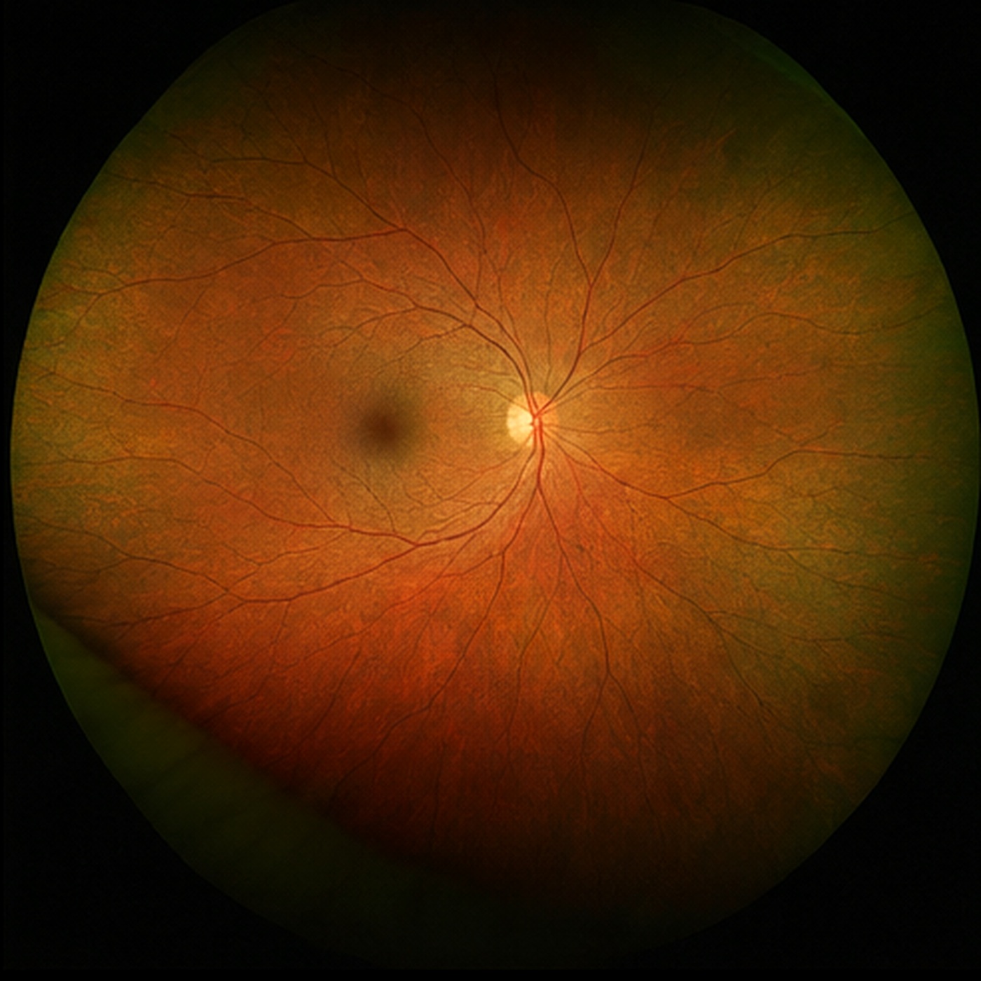
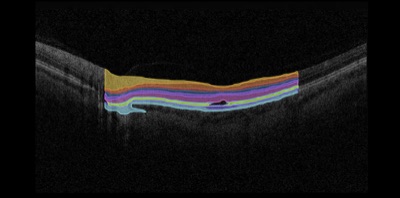
Registered widefield fundus beneath the OCT volume
Scan View
2D 3D
3D Controls
Density
Opacity
Z-Scale

Built around your day

Wherever you are in the day, your patients' images are already there.

In the exam lane Pull up a patient's full history on any computer, at any location. Nothing to install.
On the go iPhone & iPad app Made for the small screen, not shrunk from the desktop. Check a patient wherever you are.
With your patient Show them what you see: in the room, on a video visit, or in a secure message.
Dashboard
Patients Seen
284 +12.3%
Images Captured
1,247 +8.1%
Reports Generated
156 +5.7%
Avg Review Time
4.2 min -3.4%
M
T
W
T
F
S
S
OD
OD Fundus
Fundus
Dashboard
Good afternoon, Dr. Sarah Chen Lakeside Eye Care
MG
Maria Garcia 2 images · 3mo ago
DB
David Brown 2 images · 7mo ago
LT
Lisa Thompson 5 images · 9mo ago
69
Photography
Ophthalmic Ph.
Custom
OCT
Slit Lamp
Clerical
Topography
Visual Field
Other
JA
Anderson, James · #4821
48 years old
Modalities: 3 active Last visit: Oct 3
Annual visit due
Total Images 25
Avg per Visit 5.0
Images by Modality
Photography 11 images · 44%
OCT 8 images · 32%
Ophthalmic Photography 4 images · 16%
Custom 2 images · 8%
Laterality Distribution
OD
12 Right
OS
11 Left
2 Both (OU)
Device Usage
NW400
6
maestro
5
Eidon
4
iPhone 15 Pro
3
Other
2
Missing Modalities Consider capturing images for: Visual Fields, Anterior Segment
Virtual Visits
Today, Jan 29
NEXT
2:30 PM
Anderson, James Follow-up
Ready
3:15 PM
Chen, Rebecca Consultation
Waiting
4:00 PM
Patel, Arjun Screening
Scheduled
Mon
Tue
Wed
Thu
Fri
Secure Message
HIPAA
Re: Your eye exam today
SC
Dr. Chen 2:15 PM
Hi James, I've reviewed your scan from this morning. Everything looks stable compared to October.
2:18 PM James A.
Great news, thank you! Can I see it?
JA
SC
Dr. Chen 2:22 PM
Of course, here it is. We'll take another look at your follow-up in 6 months.
OCT_macula_OD.jpg Shared with you
James A.
JA
Type a secure message...
End-to-end encrypted

Hear from our community

More than 3,000 eye care professionals work in Unified every day. Here's what a few of them have to say:

"

In speaking with colleagues, I've repeatedly found I'm the only one who didn't lose any data when switching EHRs. It was all thanks to Unified Imaging. I cannot recommend it highly enough.

TH

Troy Hockemeyer, OD

Hockemeyer Eye Care

"

It's amazing how easily my Optomap data was exported to be used in an ongoing fashion. Unified Imaging is the "big red easy button" when it comes to image management.

JW

John Warren, OD

Warren Eye Care

"

Being able to review images even when out of the office is hugely helpful. Unified Imaging helps our office run efficiently; I can't imagine how anyone practices without it.

MS

Michael Samuels, OD

NC Eye Associates

"

Unified Imaging has increased my clinic efficiency. I am easily able to review patient data from all exam rooms and my office in real time. The process is completely automated.

SG

Sydnee Givre, MD, PhD

Raleigh Ophthalmology

We belong together

More for your practice, from partners who work with Unified: AI screening, at-home vision tests, exam equipment and a community of independent eye care.

Building for eye care, or running clinical research? Partner with us to reach 3,000+ eye care professionals across 1,100+ locations.

Become a Partner

Ready to get Unified?

Optimize your chair time — spend more time focusing on diagnostic data and less time managing it.

1Tell us your instruments

2We help you connect them

3Open images anywhere